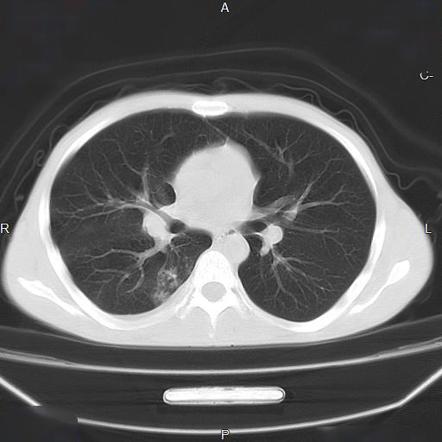
tree in bud radiology assistant
Radiology tech jobs - New York. Its microbiologic significance has not been systematically evaluated.
Chronic Airspace Disease Review Of The Causes And Key Computed Tomography Findings
Collins J Blankenbaker D Stern EJ.

. Usually somewhat nodular in appearance the tree-in-bud pattern is generally most pronounced in the lung periphery and associated with abnormalities of the. It represents dilated and impacted mucus or pus-filled centrilobular bronchioles. Tree-in-bud sign is not generally visible on plain radiographs 2It is usually visible on standard CT however it is best seen on HRCT chest.
Revision requested December 10. Tree-in-bud describes the appearance of an irregular and often nodular branching structure most easily identified in. Another important entity that can produce the tree-in-bud pattern is bronchioalveolar carcinoma BAC 1.
Of these 182 cases were excluded for the following reasons. 2 However the classic cause of tree-inbud is Mycobacterium tuberculosis especially when it. Tree-in-bud pattern simulating diffuse panbronchiolitis but without cylindrical bronchiectasis.
Tree-in-bud almost always indicates the presence of. Smooth septal thickening with basal predominance Kerley B lines ground-glass opacity with a gravitational and perihilar. Assistant bud in tree.
Irregular septal thickening usually focal or unilateral 50 adenopathy known carcinoma Reticular pattern 2. Tree in bud radiology assistant Tuesday April 12 2022 Edit. In addition Lewandowski is undoubtedly angling to get a raise something that Sport1 journalist Kerry Hau has already indicated mi Monday May.
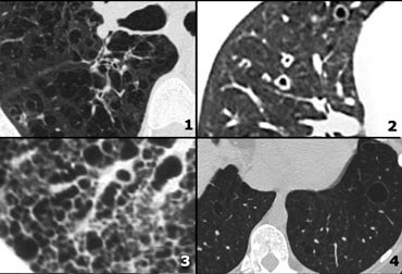
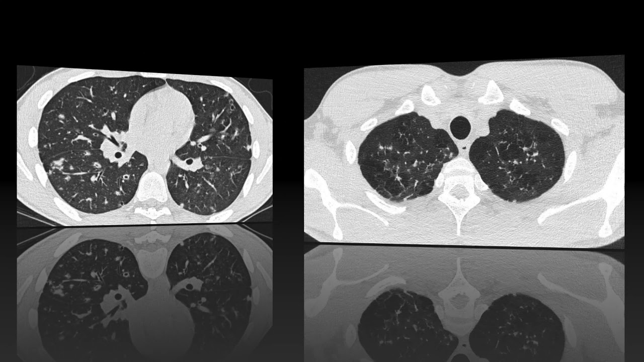
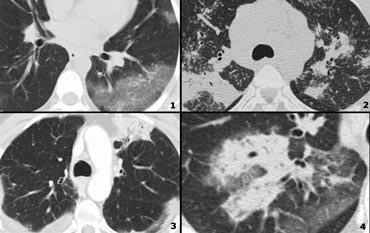
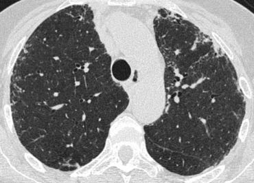
Upper and middle zone predominance. 1 refers to a pattern seen on thin-section chest CT in which centrilobular bronchial dilatation and filling by mucus pus or fluid resembles a budding tree Fig. CT patterns of.
100 died Sunday May 1 2022. The Radiology Assistant Hr Tuesday May 17 2022 Edit. 886 followed by GGA and consolidation n23.
Tree in bud radiology assistant. Tree-in-bud pattern and poorly defined nodules representing bronchiolar filling. 78 indicating the absenceresolution of TIB opacities 26 incomplete thoracic CT scan studies 75 duplicate.
The tree-in-bud appearance characterised by well-defined centrilobular nodules was observed in 1 29 patient. Mycobacterium avium complex is the most common cause in most series. Of Medicine Medical College 88 College Street Kolkata 700 073.
The most frequently observed combination of abnormalities was GGA and bronchial wall thickening n31. Typical findings of BAC on HRCT include a solitary nodule or mass 43 focal or diffuse consolidation 30 or. The connection to opacified or thickened branching structures extends proxima.
Apply For The Highest Paid Radiology tech jobs Jobs In Your Area Now. Tree-in-bud TIB is a radiologic pattern seen on high-resolution chest CT reflecting bronchiolar mucoid impaction occasionally with additional involvement of adjacent alveoli. Our Radiology Information System was searched for the term tree-in-bud from January 1 2010 to December 31 2010 iden-tifying 599 examinations.
Basic interpretation Robin Smithuis Otto van Delden and Cornelia Schaefer-Prokop Radiology Department of the Rijnland Hospital Leiderdorp and the Academical Medical Centre Amsterdam the Netherlands Secondary lobule Reticular pattern Nodular pattern Algorithm for nodular pattern Tree-in-bud. The Radiology Assistant HRCT part I. Tree in bud radiology assistant.
Tree-in-bud pattern was first described for endobronchial spread of mycobacterium tuberculosis1 It is a CT scan finding of chest with visibility of small airways. We aimed to establish the incidence of the TIB pattern as a proportion of all patients undergoing chest CT. The Tree-in-Bud Sign.
1 From the Department of Radiology University of Vienna Waehringer Guertel 18-20 A-1090 Vienna Austria. Primary pulmonary lymphoma or leukemia 247. Tree-in-bud appearance represents dilated and fluid-filled ie.
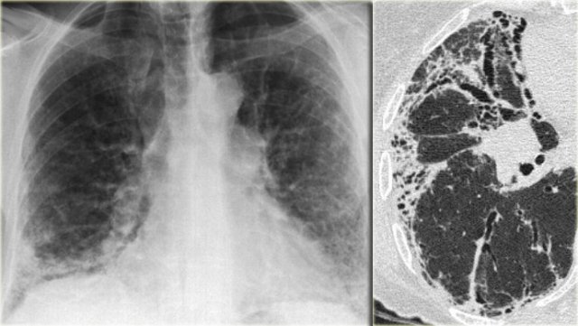
Endobronchial spread of infection TB MAC any bacterial bronchopneumonia Airway disease associated with infection cystic fibrosis bronchiectasis less often an airway disease associated primarily with mucus retention allergic bronchopulmonary aspergillosis asthmaDiagnosis Radiology. Tree in bud radiology assistant Wednesday May 18 2022 Edit. Typically the centrilobular nodules are 2-4 mm in diameter and peripheral within 5 mm of the pleural surface.
Small nodules some related to vessels admixed with ground-glass opacity and consolidation. Study with Quizlet and memorize flashcards terms like Reticular pattern 1. Received November 11 1999.
It consists of small centrilobular nodules of soft-tissue attenuation connected to multiple branching linear structures of similar caliber that originate from a single stalk. Revision received and accepted May 22 2000. The tree-in-bud pattern is commonly seen at thin-section computed tomography CT of the lungs.
Memorial celebration of life on Saturday May 21 2022 at 11 am at Venice Church of the Nazarene 1535 E May 18 2022 Edit. CTHRCT high-resolution-CT showed extensive bronchiectasis along with parenchymal disruption i Wednesday May 18 2022 Edit. Tree in bud opacification refers to a sign on chest CT where small centrilobular nodules and corresponding small branches simulate the appearance of the end of a branch belonging to a tree that is in bud.
Tree In Bud Sign Lung Radiology Reference Article Radiopaedia Org. 657 and bronchial wall thickening and consolidation n22. Address correspondence to the author e-mail.
3 found that the tree-in-bud pattern was seen in 256 of the CT scans in patients with bronchiectasis. Originally reported in cases of endobronchial spread of Mycobacterium tuberculosis this. Tree-in-bud describes the appearance of an irregular and often nodular branching structure most easily identified in the lung periphery.
31 March 2013. Apply Today Start Tomorrow. Originally and still often thought to be specific to endobronchial Tb the sign is actually non-specific and is the.
Assistant Professor Dept. Assistant bud in tree. This pattern is manifested by luminal filling of contiguous branching segments of bronchioles seen in bronchiolar disease.
In centrilobular nodules the recognition of tree-in-bud is of value for narrowing the differential diagnosis. Memorial celebration of life on Saturday May 21 2022 at 11 am at Venice Church of the Nazarene 1535 E. As the technique calls for 1-2 mm slice.
100 died Sunday May 1 2022.
Tree In Bud Sign Lung Radiology Reference Article Radiopaedia Org

Reversed Halo Sign Lungs Radiology Reference Article Radiopaedia Org
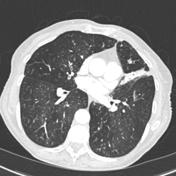
Tree In Bud Sign Lung Radiology Reference Article Radiopaedia Org
Pin De Gerardo Valenzuela En Neuroimagen

Learningradiology Lung Abscess Pulmonary Lunges Pulmonary X Ray

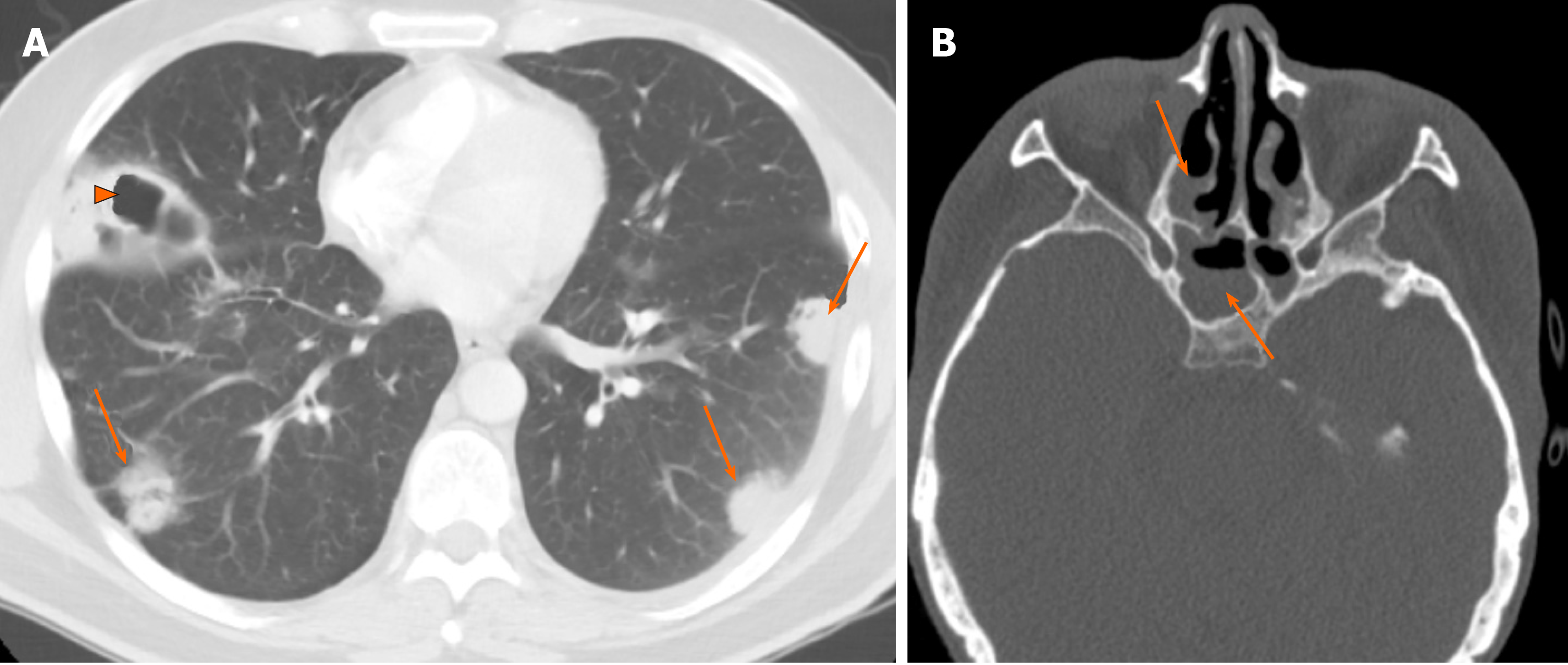
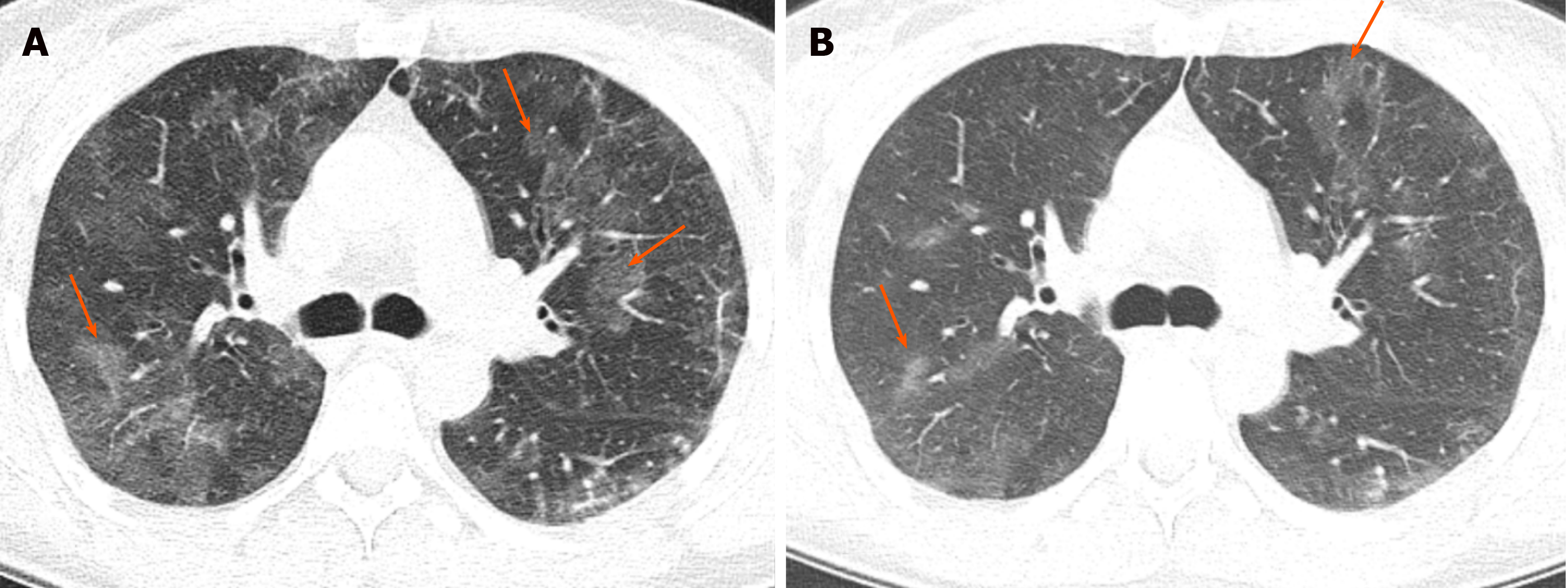
A And B Axial Hrct Cuts Shows Centrilobular Nodules With Tree In Download Scientific Diagram

Tree In Bud In Centrilobular Nodules The Recognition Grepmed
The Radiology Assistant Hrct Basic Interpretation
2
The Radiology Assistant Hrct Basic Interpretation
The Radiology Assistant Hrct Common Diagnoses
The Radiology Assistant Hrct Common Diagnoses

Tree In Bud Sign Lung Radiology Reference Article Radiopaedia Org
The Radiology Assistant Hrct Basic Interpretation
The Radiology Assistant Hrct Basic Interpretation

Case 4 2021 A 70 Year Old Woman With Dyspnea On Exertion And Abnormal Findings On Chest Imaging Nejm

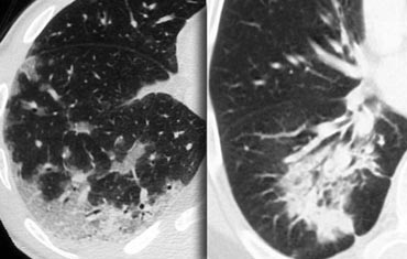
Cylindrical Bronchiectasis And Tree In Bud Pattern In Lower Lobes And Download Scientific Diagram
Chronic Airspace Disease Review Of The Causes And Key Computed Tomography Findings

Tree In Bud Almost Always Indicates The Presence Of Endobronchial Grepmed